Canine Meniscus Assessment and Treatment
Understanding Dog Meniscus Anatomy
The canine meniscus plays a crucial role in a dog’s knee joint health, providing both stability and shock absorption. Each knee joint in a dog contains two menisci: the medial meniscus and the lateral meniscus. These cartilage structures work as cushions between the bones, absorbing impact and distributing weight across the knee joint to protect the bone surfaces. The meniscus is especially vulnerable to injury in dogs with cranial cruciate ligament (CCL) ruptures, which often leads to a torn meniscus in dogs, specifically in the medial meniscus.
Normal Dog Meniscus
There are two menisci (the medial meniscus and the lateral meniscus) in each dog knee that serves as cartilage cushions or shock absorbers.
Due to this shock absorbing effect, a healthy canine meniscus helps prevent the dog’s knee joint from wear and tear. In dogs with CCL injuries, the medial meniscus is often affected, while the lateral meniscus is rarely torn. Proper treatment of dog meniscus injuries following a CCL rupture includes thoroughly evaluating the menisci to ensure there is no tear.
Arthroscopy is one of the best methods to assess dog knee injuries, offering a minimally invasive and accurate way to detect tears. If a canine meniscus tear is found during the arthroscopy, the damaged portion is removed or, in rare cases, repaired.

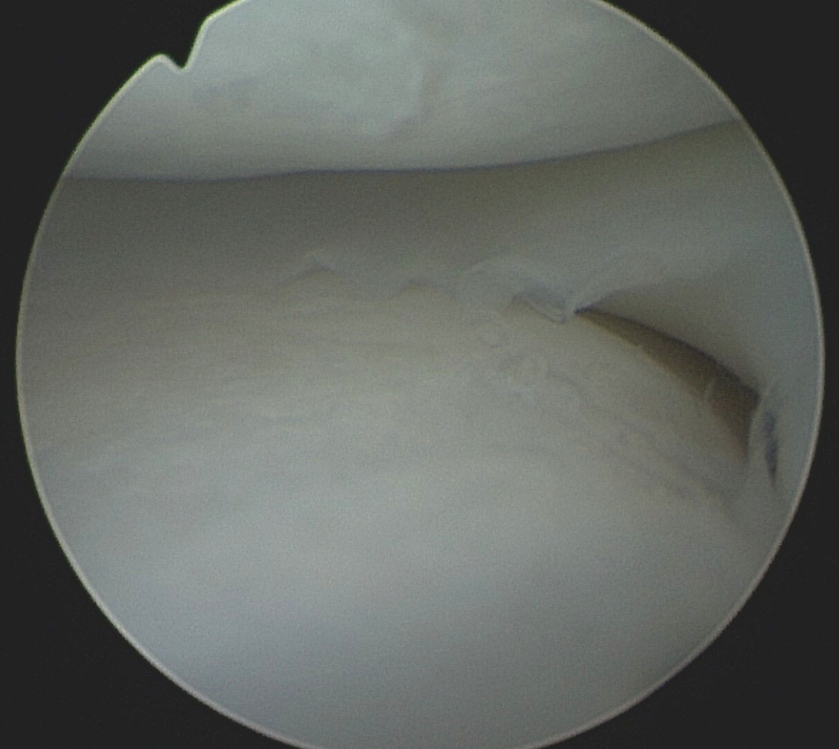
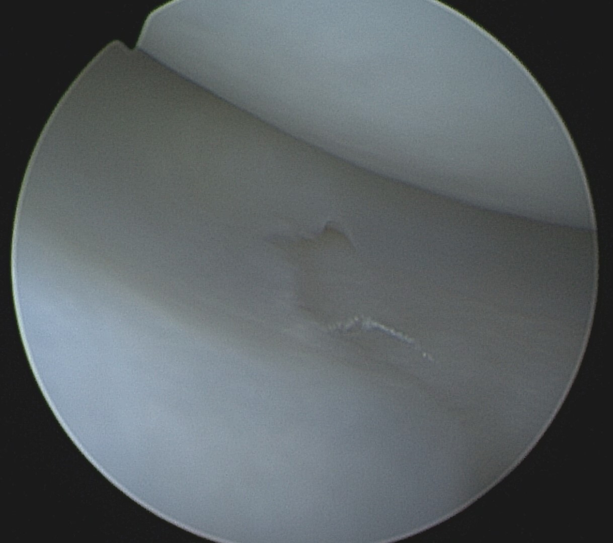
The following images and video demonstrate a normal canine medial meniscus. Special thanks to Dr. Sam Franklin and the University of Georgia Educational Resources Center for their collaboration in capturing these educational resources.
Torn Dog Meniscus
A torn meniscus in dogs is typically characterized by the back portion of the meniscus displacing forward between the femur and tibia. This can sometimes (rarely) be heard by owners as a clicking or popping sound, referred to as a meniscal click. In the arthroscopic video below, veterinary surgeon Dr. Sam Franklin demonstrates a dog meniscus tear where the meniscus displaces forward repeatedly when the tibia moves due to the CCL rupture. The video illustrates how pushing the torn meniscus back into place won’t resolve the issue. This torn portion of meniscus needs to be removed or, in rare cases, repaired.
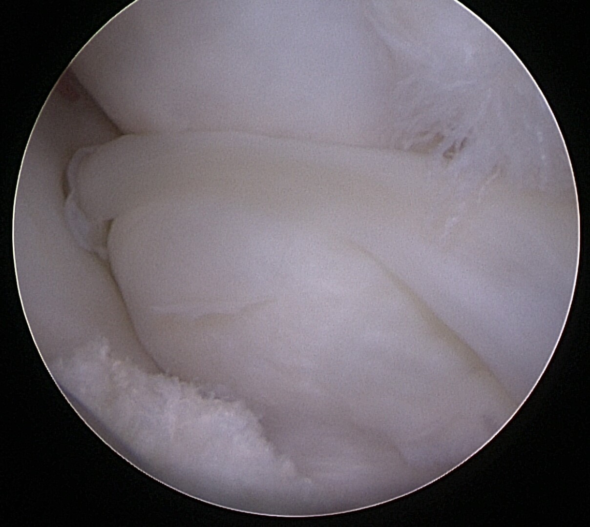
In many cases the torn meniscus become permanently wedged between the femur and the tibia as shown in the image below top, right. This is an arthroscopic image of a whole back half of a medial canine meniscus that is torn and displaced forward sitting between the femur and tibia, similar to the video on the left. This is referred to as a peripheral detachment and the dog’s torn meniscus needs to be removed.
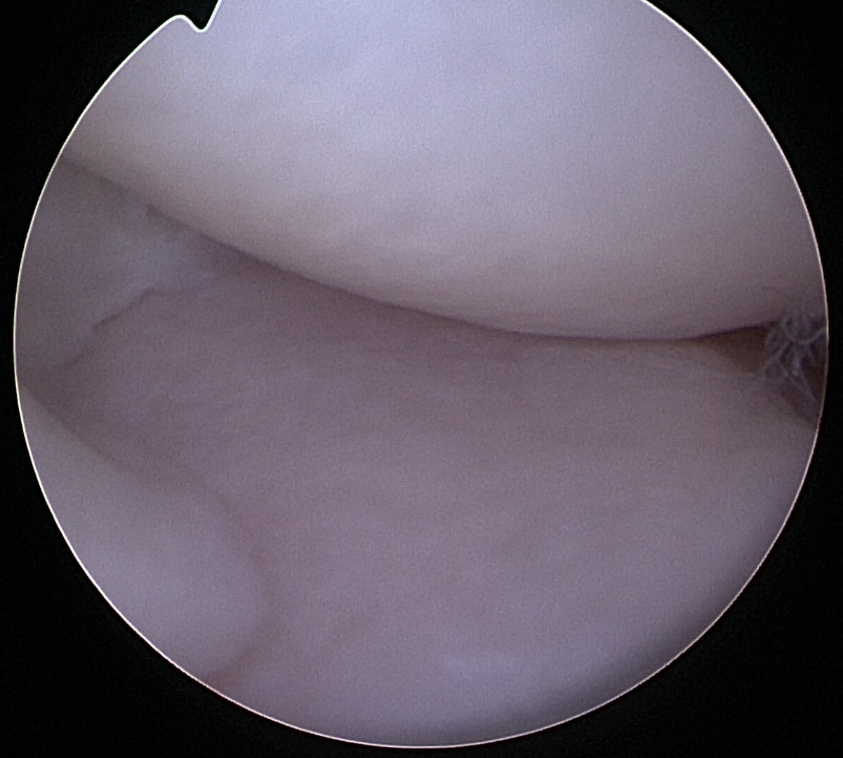
Following removal of the torn portion of the canine meniscus the remaining meniscus can be seen (within the left side of the image to the bottom right) and the femur (in the upper portion of the image to the bottom right) can now comfortably rest on the tibia. Note that the cartilage in this dog’s knee is still in good health and removal of the meniscus does not equate to “bone on bone” contact.

How to Treat a Torn Meniscus in Dogs
To treat dogs with a torn meniscus, our team recommends arthroscopy, a minimally invasive surgical procedure used to diagnose and treat joint problems in dogs. It involves inserting a small camera (arthroscope) into the joint through tiny incisions, allowing our skilled veterinary surgeons to see the joint’s interior without needing to perform a large open surgery. By addressing the dog’s torn meniscus and any related CCL issues during a single procedure, we help ensure optimal recovery and mobility.
Check out our page on arthroscopy to learn more about how it can help diagnose and treat dog meniscus injuries effectively.

