ANGULAR LIMB DEFORMITIES
Angular limb deformities are relatively common in dogs and manifest as a leg that is not straight. There are 3 potential causes of limb deformities:
-
Damage to a growth plate during the period of skeletal growth.
-
Abnormal development of a limb during skeletal growth in dogs for whom we have selected them to have abnormal skeletal conformation and who are genetically predisposed to abnormal growth. Such breeds of dogs are referred to as chondrodystrophic breeds (meaning abnormal cartilage development) and include breeds like Shih-Tzus, Dachsunds, Bassett Hounds, and other dogs that usually have short and malangulated limbs.
-
Fracture of a limb that heals with poor alignment, referred to as a malunion.
Of those potential causes of angular deformity, the most common causes are damage to a growth plate in an otherwise normal dog or abnormal development in a chondrodystrophic breed. Malunion, secondary to fracture, is less common.
Of the deformities that we see, malangulation of the lower forelimb (lower front leg) is the most common. Dogs have two bones in their forelimb, just as we humans do, the radius and the ulna. As a dog grows these two bones must develop synchronously. Commonly, the growth plate of the distal ulna (adjacent to the wrist) becomes injured because its shape and location predispose it to injury. As a result of the injury the ulna stops growing but the radius continues to grow. In such cases the ulna hinders the growth of the radius, typically resulting in an abnormal curvature of the lower forelimb.
TREATMENT OF LIMB DEFORMITIES
The ultimate goals of deformity correction are to straighten (re-align) the limb and there are many different methods by which this can be done. However, the first and often crucial step in treating a limb deformity is planning the correction accurately. This is crucial because every deformity is unique and requires a unique correction.
The traditional method of evaluating and planning deformity correction is to make radiographs (take X-ray) and to evaluate and plan surgical correction accordingly. Such method is simple, relatively inexpensive, and often effective, particularly for relatively simple deformities.
However, there are some limitations to using radiography (X-rays) for assessment of the deformity and planning of correction. The greatest limitation is that accurately characterizing the deformity in 3 dimensions can be limited when using 2-dimensional images (i.e. X-rays). This is particularly true if the deformity is complex and involves malangulation in all 3 planes of space.
Thanks to advances in cross sectional imaging technology, 3-dimensional (3D) modeling, and 3D printing, we can now more accurately characterize, replicate, print, and plan our surgeries for complex ALDs in 3 dimensions.
The process involves performing a CT scan of the dog’s limb. Thanks to advances in CT imaging this is quick and easy. Dogs are briefly sedated and a CT scan of the limb is performed. The information is then exported from the CT scanner to a work station with 3D modeling software that facilitates turning such information into a 3D rendering of the limb. This process can take minutes to hours depending upon the skill level of the person performing the rendering and the complexity of the deformity.
Once the 3D rendering is created we can visualize and manipulate the limb in 3 dimensions. Such process can improve our planning for surgery and often is sufficient to enable accurate planning at this stage. However, we can take the process a step further. These 3D renderings of the limb can be used to guide 3D printing of the limb such that we have physical replicas of the limb.

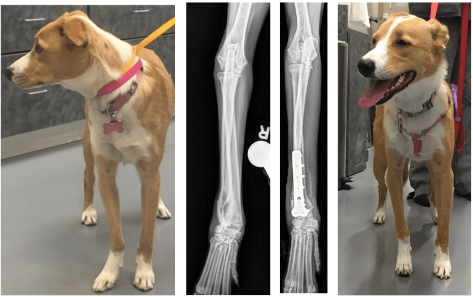
Patient with a relatively simple deformity of the right forelimb that was characterized with radiography (i.e. X-ray), fixed with a bone plate in screws, and had an excellent outcome.
CORRECTIVE OSTEOTOMY
All treatment of ALDs requires performing a corrective osteotomy. This means we need to cut the bone (osteotomy) to allow re-alignment and straightening. The actual straightening of the limb can be done acutely, which means that we re-align the limb in surgery such that it is straight immediately following surgery. This is the most common way of treating deformities in dogs. However, in some dogs with complex deformities we perform re-alignment gradually over a period of several days using an external skeletal fixator. The greatest advantage of slow distraction and re-alignment is that gradual distraction can enable bone growth and lengthening in short limbs, rather than just straightening of the limb.

Sissy prior to correction of her angular limb deformities. A) 3D virtual rendering of the right forelimb of Sissy. B) 3D printed model of Sissy’s left forelimb C) An osteotomy guide is then made to control/guide where the osteotomy will be made. D) The surgery is then rehearsed on the model of Sissy’s forelimb. E) Completion of the rehearsal surgery ensures that the osteotomy guide and planned oblique plane osteotomy will result in optimal re-alignment of the limb. The osteotomy guide can then be removed, sterilized, and used during surgery to ensure exact replication of the surgery that was rehearsed on the model. Sissy after correction of her forelimb deformities with excellent results in the realignment of the forelimb.
FIXATION OF ANGULAR LIMB DEFORMITIES

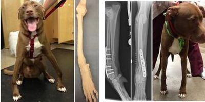
Images (from left to right) of a dog with deformity of both forelimbs, one of the limb models made following CT, post-operative X-rays showing straight a limb that is stabilized with a bone plate and screws, and a post-operative photograph showing excellent re-alignment of both limbs.
BONE PLATES AND SCREWS
Bone plates and screws can be used when acute correction of a deformity is to be performed. One advantage of bone plates and screws is that they are relatively easy to apply for an experienced surgeon. They are also very strong and tend to be very effective in keeping the bone stable as the bone heals. They are also easy to manage post-operatively because there is nothing for owners or surgeons to do once they are placed because they are entirely within the body. As a result, bone plates and screws are very commonly used for the correction of deformities.
The disadvantages of bone plates are that should they become infected a minor surgery may be required in the future to remove them once the bone is healed. However, the likelihood of infection of bone plates is small and the more relevant disadvantage of bone plates is that they cannot be ‘adjusted’ to slightly improve alignment of the bones once the surgery is completed. The disadvantages explain the value of investing time and energy in meticulous surgical planning, including potentially using CT scans, 3D modeling and 3D printing prior to surgery, and coupling these with acute correction and use of bone plates and screws. Such overall approach to management of deformities can be appealing because it means that the osteotomy is stabilized with a bone plate and screws which is easy for the owner and dog post-operatively. And, the disadvantage of the bone plate and screws (inability to change alignment after surgery is over) is mitigated by planning and maximizing the likelihood that alignment will be perfect post surgery.
EXTERNAL SKELETAL FIXATORS (ESF)
External skeletal fixators includes placement of pins or wires through small incisions in the skin and into the bones. These pins and wires are then all connected together outside the body to hold the different segments of bone in their new and optimal alignment. The advantages of ESF include that one can make a small incision through which the osteotomy is performed (the bone is cut) and then very small incisions through which the pins or wires are placed. Therefore, in total, the size and total length of incisions can be reduced and the surgery can be performed in a less invasive manner.
Another appealing feature of ESFs is that they provide some ability to change or optimize alignment after surgery is performed. Because the pins and wires that are inserted into the bone are all connected to each other outside the body, if the alignment of the bone is not perfect one can re-align the pins and wires that are connected to the bone and thus re-align the bone segments. Sometimes this is particularly appealing if post-operative X-rays show good alignment but also demonstrate that alignment could still be further improved. It is this general principle of being able to manipulate the segments of bone from outside the body after surgery that also allows planned, gradual distraction over time to both lengthen and straighten a bone over time.
The disadvantages of ESF are that it is more labor intensive to manage following surgery for both the owner and the surgeon and there is more commonly morbidity associated with minor pin tract infections. This means that the areas where the pins penetrate the skin can become infected. In an effort to mitigate this possibility, the owner and surgeon typically spend time cleaning these areas at least weekly, if not daily, to try and decrease the likelihood of infection. However, despite such protective cleaning, pin tract infection is still more likely than infection with bone plates and screws that are placed entirely within the body. This is because the pins/wires traverse the skin, which is the dog’s greatest barrier to infection. The good news is that once the bone is healed, an external skeletal fixator is removed with sedation; with such removal, infection is typically resolved.
CHOOSING BONE PLATE VS EXTERNAL SKELETAL FIXATOR
The choice between using ESF or bone plates to stabilize a corrective osteotomy is a decision that the surgeon makes on an individual basis. As stated above, every deformity is unique and so each case has to be considered on an individual basis.
PRE-OPERATIVE IMAGING
A thorough and accurate evaluation of a deformity using X-rays often requires multiple, perfectly positioned and acquired X-rays. Hence, owners should be prepared for dogs to be briefly sedated for such process. A CT scan may be recommended to evaluate and characterize the deformity. A CT will enable Kansas City Canine Orthopedics to create 3D models and/or virtually plan the surgery.
Veterinary Services
Below are all of the veterinary services we offer at Kansas City Canine Orthopedics. If you have any questions regarding our services, please feel free to call us.

